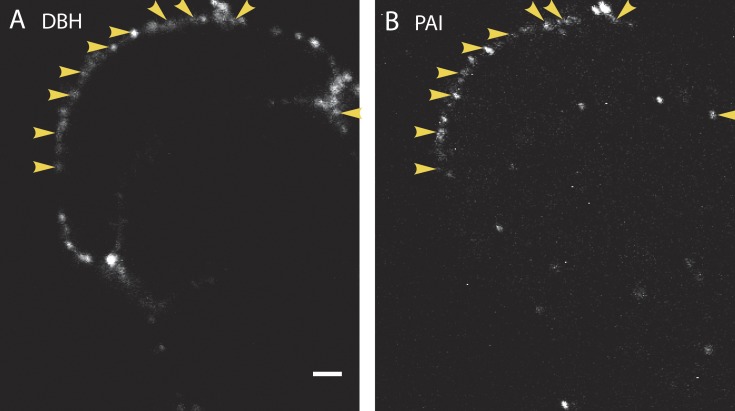
Figure 2.
Endogenous PAI colocalizes with DBH on the cell surface after stimulation. Cultured bovine chromaffin cells were stimulated for 10 s with 56 mM K+ at 34°C. The solution was replaced with buffer containing 5.6 mM K+, and the cells were immediately placed on ice. Cells were then incubated with antibodies to PAI (B) and to the lumenal domain of the granule membrane protein DBH (A) for 60 min on ice, and then processed and imaged by confocal microscopy. Because the cells were not permeabilized, only antigens present on the surface of the cells are visible. Arrowheads indicate instances of colocalization of secreted DBH and PAI. n = 10 cells. Bar, 2 µm.

