
Figure 3. The 4.5S ribosomal RNA and its interactions with ribosomal proteins.
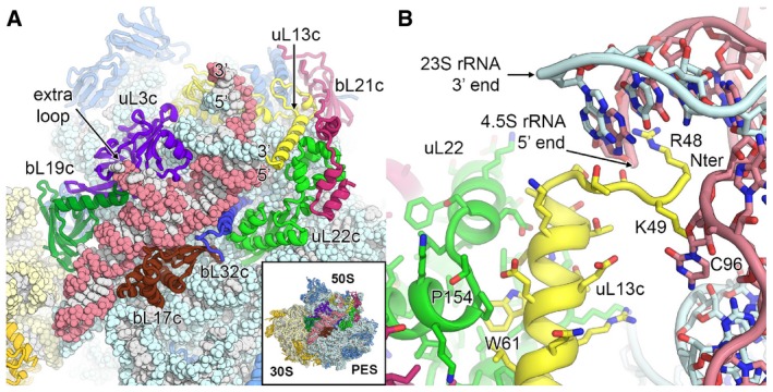
- Position of the 4.5S rRNA (red) at the surface of the 50S large subunit. The 3′ and 5′ ends of the 4.5S rRNA and the 23S rRNA (blue) are labelled. Ribosomal proteins interacting with or in close proximity to the 4.5S rRNA are shown in different colours.
- Stabilization of the 5′ end of the 4.5S rRNA and the 3′ end of the 23S rRNA by the plastid‐specific N‐terminal tail of uL13c (yellow). Specific residues of uL13c, uL22c and 4.5S rRNA are labelled.