
Figure 4. Architecture of the mRNA entry and exit site.
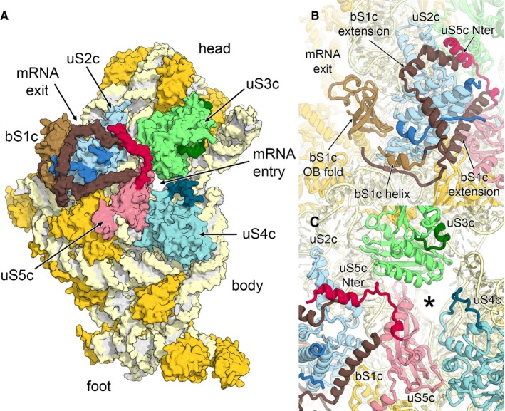
- Surface representation of the solvent accessible side of the 30S small subunit. Ribosomal proteins bS1c (brown), uS2c (blue), uS3c (green), uS4c (cyan) and uS5c (red) are labelled, and plastid‐specific elements are indicated by darker colour shades.
- Helical extensions of bS1c cluster around uS2c.
- Chloroplast‐specific extensions of uS4c and uS5c remodel the mRNA entry site (marked with an asterisk).