
Figure 5. Architecture of the polypeptide tunnel exit.
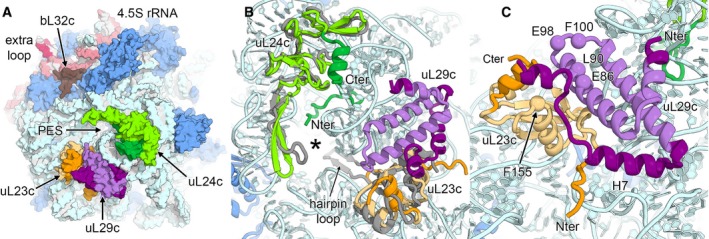
- Surface representation of the polypeptide exit site (PES) of the 50S large subunit. Ribosomal proteins located around the PES are uL23c (orange), uL24c (green) and uL29c (violet). Plastid‐specific features are indicated by darker colour shades.
- Structural differences of the PES (marked with an asterisk) of the chloroplast 50S in comparison with the bacterial 50S subunit. The bacterial ribosomal proteins uL23 and uL24 (both in grey) are overlaid.
- Adaptations of putative binding sites of cpSRP54. Corresponding residues involved in NG‐domain binding in the bacterial ribosome are shown as spheres and are labelled.