
Figure 6. Plastid translation factor pY.
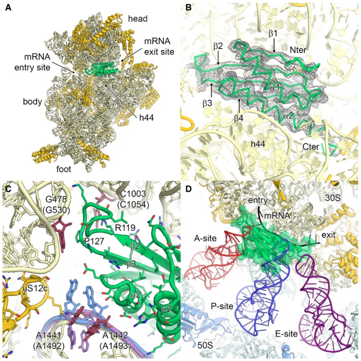
- Binding of plastid translation factor pY, shown in lime green, to the mRNA channel of the small subunit. The small subunit is shown from the intersubunit side. The 16S rRNA is coloured in pale yellow, and ribosomal proteins are in gold.
- EM density for plastid pY. Secondary structure elements and N‐ and C‐termini are indicated.
- Molecular interaction of plastid pY with 16S rRNA. The conserved nucleotides involved in A‐site decoding are coloured in red and labelled (bacterial numbering in brackets). The bacterial nucleotides A1492 and A1493 of the empty 30S subunit (PDB 1J5E; Wimberly et al, 2000) and of the 70S ribosome in complex with mRNA and tRNAs (PDB 4V51; Selmer et al, 2006) are overlaid and coloured in purple and blue, respectively. Pro127 and Arg119 of plastid pY are indicated.
- Superposition of the chloroplast 70S:pY complex with bacterial A‐, P‐ and E‐site tRNAs and mRNA from the crystal structure of the Thermus thermophilus 70S ribosome (PDB 4V51; Selmer et al, 2006).