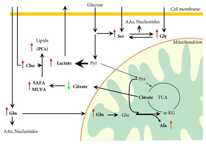
Figure 2.
Metabolomic alterations and active metabolic pathways in proliferating thyroid cancer cells. Metabolomic analysis of thyroid cancer (TC) tissues indicates increased glycolytic rate, lactate production, glutaminolysis, and lipid biosynthesis. TC cells upregulate glucose uptake through the overexpression of the glucose transporter 1 (GLUT1). The pyruvate generated by the glycolysis is diverted toward lactate production through the action of lactate dehydrogenase A (LDH-A), overexpressed in TC and kept away from mitochondrial oxidative metabolism. Lactate is exported from tumor cells via a monocarboxylate transporter. The extracellular lactate activates the angiogenesis and provides metabolic fuel to proliferating cancer cells. The increase of serine and glycine may be explained by either increased uptake via neutral amino acid transporters or de novo biosynthesis of serine from the glycolytic intermediate 3-phosphoglycerate. Serine can be converted to glycine by the action of serine hydroxymethyltransferase that shows higher expression rate in TC. Therefore, de novo serine biosynthesis sustains glycine biosynthesis. The so-called “one carbon metabolism,” including both serine and glycine, cycles carbon units from amino acids to support amino acid and purine biosynthesis. Glutamine, also involved in amino acid and nucleotide biosynthesis, is taken up into the cell through the glutamine importer ASCT2 and is deaminated in mitochondria by glutaminase to form glutamate. Glutamate dehydrogenation produces alpha-ketoglutarate to replenish the tricarboxylic acid cycle intermediates. The enzymes involved in glutaminolysis have been found overexpressed in TC. It is still unclear whether the glutamate transamination with pyruvate to alanine and alpha-ketoglutarate, recently demonstrated in colon cancer cells, occurs in TC. Conversion of alpha-ketoglutarate to citrate supports the biosynthesis of cytosolic acetyl-CoA and, then, of saturated and monounsaturated fatty acids. Accordingly, increased expression levels of fatty acid synthase and desaturases have been found in TC. The increase of choline levels and the activation of fatty acid biosynthesis fuel the biosynthesis of lipids such as phosphatidylcholines. Green and red arrows indicate lower and higher metabolite levels, respectively, in TC compared with nontumor lesions or adjacent normal tissue. AAs: amino acids; Ala: alanine; α-KG: alpha-ketoglutarate; Cho: choline, Gln: glutamine; Glu: glutamate; Gly: glycine; MUFA: monounsaturated fatty acids; PCs: phosphatidylcholines; Pyr: pyruvate; SAFA: saturated fatty acids; Ser: serine; TCA: tricarboxylic acid cycle.

