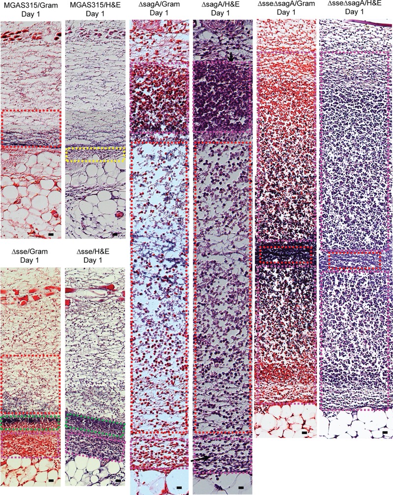
FIG 4.
Histological analyses of skin infection sites infected with MGAS315 and the Δsse, ΔsagA, and Δsse ΔsagA mutants in mice at day 1 after inoculation. Groups of 10 6-week-old female C57BL/6J mice were subcutaneously inoculated with 1.5 × 10⁸ CFU of MGAS315, 1.8 × 10⁸ CFU of the Δsse mutant, 1.9 × 10⁸ CFU of the ΔsagA mutant, or 2.0 × 10⁸ CFU of the Δsse ΔsagA mutant in 0.2 ml DPBS. Five mice from each group were sacrificed on days 1 and 2 after inoculation to collect the skin infection sites. The skin infection sites were fixed and analyzed with Gram and H&E stains, as described in Materials and Methods. Shown are representative Gram- and H&E-stained images of the regions in the skin infection sites indicated by the boxes in Fig. S1, S3, S5, and S7 in the supplemental material. Bars, 20 μm.

