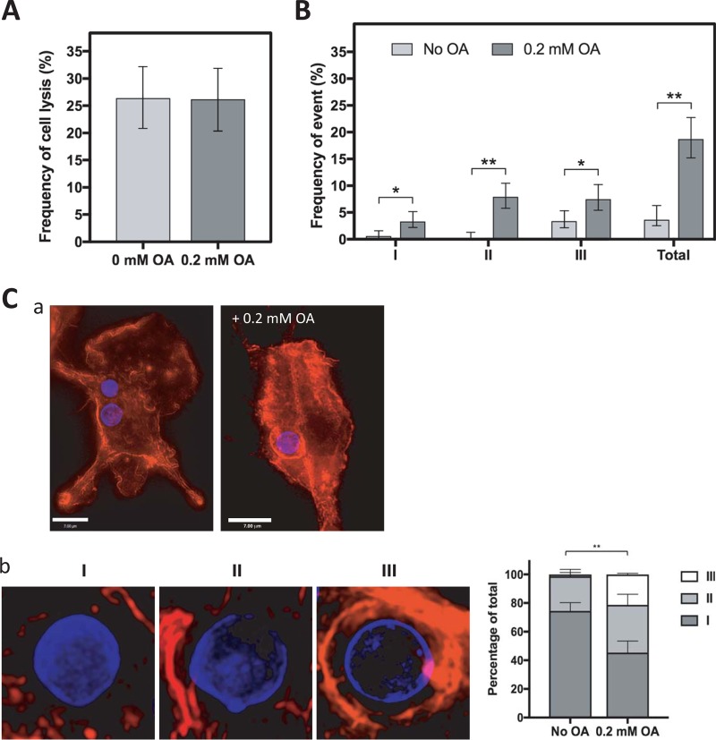
FIG 6.
Effect of OA on the mode of C. neoformans egress. (A) The frequency of cell lysis was counted from 24-h time-lapse movies of BMDM treated with or without 0.2 mM OA and infected with C. neoformans. Experiments were performed in triplicate. Error bars represent the 95% confidence interval of the mean. (B) BMDM were treated with or without 0.2 mM OA during a C. neoformans infection. The frequencies of nonlytic exocytosis (NLE) events (type I, complete NLE; type II, partial NLE; type III, cell-to-cell transfer) were counted from 24-h time-lapse movies. Experiments were performed in duplicate. Error bars represent the 95% confidence interval of the mean. *, P < 0.01; **, P < 0.0001 by Fisher's exact test. (C) Fluorescence microscopy of Uvitex-labeled C. neoformans infecting BMDM, 4 h postinfection, stained with Alexa594 –F-actin. (a) A more pronounced actin staining is visible surrounding the C. neoformans-containing phagosome when incubated with 0.2 mM OA. Scale bar, 7 μm. (b) Class distribution of the actin coverage surrounding the C. neoformans-containing phagosome with 0.2 mM OA or no OA (control): little to none (0 to 30%; I), half (30 to 70%; II), and full (70 to 100%; III). **, P < 0.0001 by two-way ANOVA with standard deviations shown, Tukey's multiple-comparison test.

