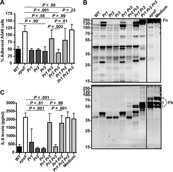
FIG 2.

Predominant contribution of StmPr1 to Xps-mediated activities. (A) A549 cells were incubated for 3 h with 25% (vol/vol) culture supernatant collected from the WT, xpsF mutant NUS4, stmPr1 mutant NUS5 (Pr1), stmPr2 mutant NUS6 (Pr2), stmPr3 mutant NUS11 (Pr3), stmPr1 stmPr3 mutant NUS12 (Pr1 Pr3), stmPr2 stmPr3 mutant NUS13 (Pr2 Pr3), stmPr1 stmPr2 mutant NUS7 (Pr1 Pr2), or stmPr1 stmPr2 stmPr3 mutant NUS14 (Pr1 Pr2 Pr3). Cell detachment relative to that for control cells treated with medium alone was determined as described in the legend to Fig. 1. (B) Human fibronectin (Fn; top) and fibrinogen (Fb; bottom) were incubated at 37°C for 16 h with 25 μl of culture supernatant from the WT, the xpsF mutant, the indicated protease mutant strains, or a control treated with medium alone. ECM protein degradation was analyzed as described in the legend to Fig. 1. The fibrinogen α, β, and γ chains are indicated. The migration of molecular mass standards (in kilodaltons) is indicated to the left of the gel images. Although the samples were examined on the same gel, they were not in adjacent lanes, and therefore, we cropped out intervening lanes that were not pertinent to the analysis. Data are representative of those from three independent experiments. (C) Recombinant IL-8 was incubated at 37°C for 16 h with 50 μl of culture supernatant collected from the WT strain, the xpsF mutant, the indicated protease mutant strains, or a control treated with medium alone. IL-8 levels were quantified by ELISA. For panels B and D, data are represented as the mean and SD from three independent experiments.
