FIG 3.
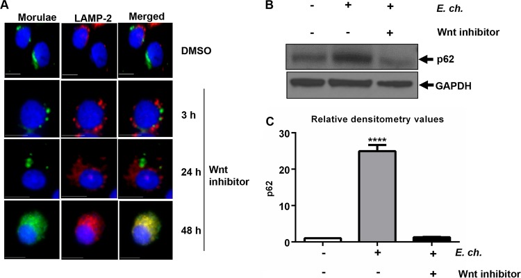
E. chaffeensis-mediated inhibition of phagolysosome fusion and p62 degradation depends on the Wnt signaling pathway. E. chaffeensis-infected THP-1 cells (24 h) were treated with either DMSO or Wnt inhibitor (3289-8625; 30 μM). (A) Cells were harvested at 3, 24, or 48 h posttreatment, fixed, permeabilized, and probed with polyclonal anti-E. chaffeensis (green) and anti-LAMP2 (red) antibodies. Colocalization of ehrlichial vacuole with lysosome (LAMP-2) was observed at 48 h posttreatment with Wnt inhibitor. Cells were visualized by immunofluorescence microscopy (×40; bars, 10 μm). Cell nuclei were stained with DAPI (blue). (B) Immunoblotting of cell lysates from infected or uninfected THP-1 cells (24 h) after treatment with Wnt inhibitor for 48 h. Membranes were probed with antibodies against p62 and GAPDH to measure lysosomal degradation. Data are representative of results from n = 4 experiments. (C) Relative band intensities of p62 normalized to the loading control (GAPDH) were determined using ImageJ software (****, P < 0.0001 [Student's t test]; n = 3).

