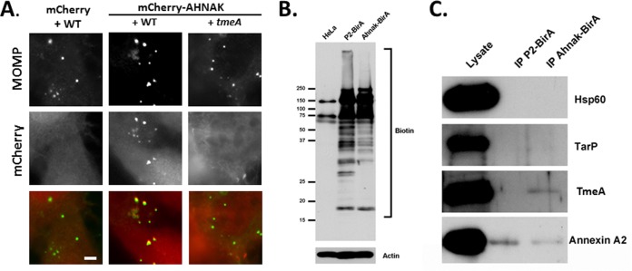
FIG 6.
Association of AHNAK and TmeA during chlamydial invasion. (A) HeLa cells were nucleofected with pmCherry or pmCherry-Ahnak. At 24 h cells were infected at 4°C with C. trachomatis L2 WT or tmeA strain at an MOI of 20. Cultures were shifted to 37°C for 30 min and subsequently were fixed with paraformaldehyde and processed for direct and indirect immunofluorescence. Chlamydia organisms were detected using anti-MOMP (green). mCherry or mCherry-Ahnak (red) is shown in the mCherry channel. Scale bar, 5 μm. (B) HeLa cells or cells expressing P2-BirA or AHNAK-BirA were cultivated in the presence of biotin for 15 h, and whole-culture material was resolved via SDS-PAGE. Material was probed with NeutrAvidin-HRP to visualize biotin- or actin-specific antibodies as a loading control. Positions of molecular size markers are indicated. (C) Biotinylated proteins were immunoprecipitated (IP) from cleared lysates of cultures infected for 5 h and expressing P2-BirA or AHNAK-BirA. Material from cleared lysates (lysate) or immunoprecipitates was probed with Hsp60-, TarP-, TmeA-, or annexin A2-specific antibodies in immunoblots and visualized via chemiluminescent detection.

