Figure 3.

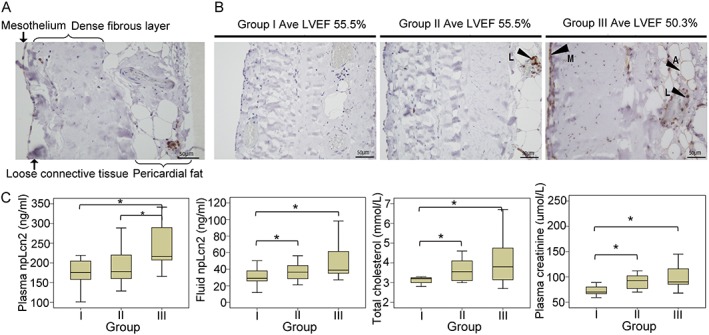
Expression and distribution of non‐polyaminated lipocalin‐2 (npLcn2) protein in the pericardium tissue biopsies collected from Danish patients during cardiothoracic surgery. (A) Immunohistochemical staining was performed for pericardium tissue sections using polyclonal antibodies against npLcn2. (B) Samples were stratified by the pattern of npLcn2 expression and distribution in pericardium tissue sections. Note that the average left ventricular ejection fraction (LVEF) of patients in Group III was significantly lower than that of Groups I and II. Arrows indicate the positive staining of npLcn2. A = adipocytes, M = mesothelial cells, and L = leucocytes. Magnification, ×200. (C) Based on the number of positively stained cells and the distribution pattern, samples were separated into three groups for comparison of their lipocalin‐2, total cholesterol, and plasma creatinine levels. *P < 0.05 (n = 9–12).
