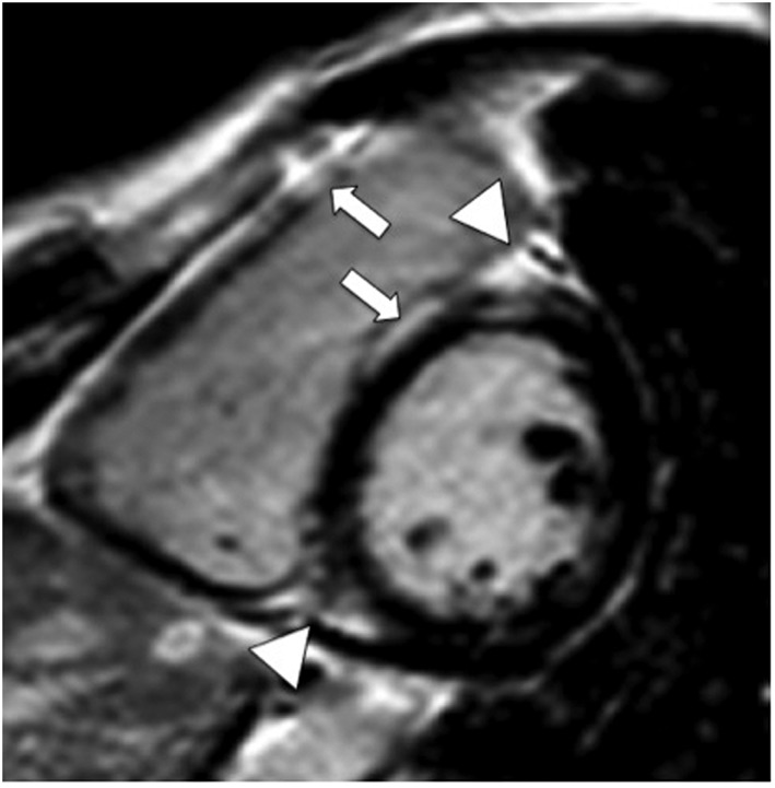
Figure 2.

Contrast‐enhanced magnetic resonance study (inversion‐recovery gradient echo sequence, end‐diastolic frame, and short axis view) demonstrates enhancement of the right ventricular free wall (arrow), ventricular insertion points (triangles), and right‐sided septum (arrow).
