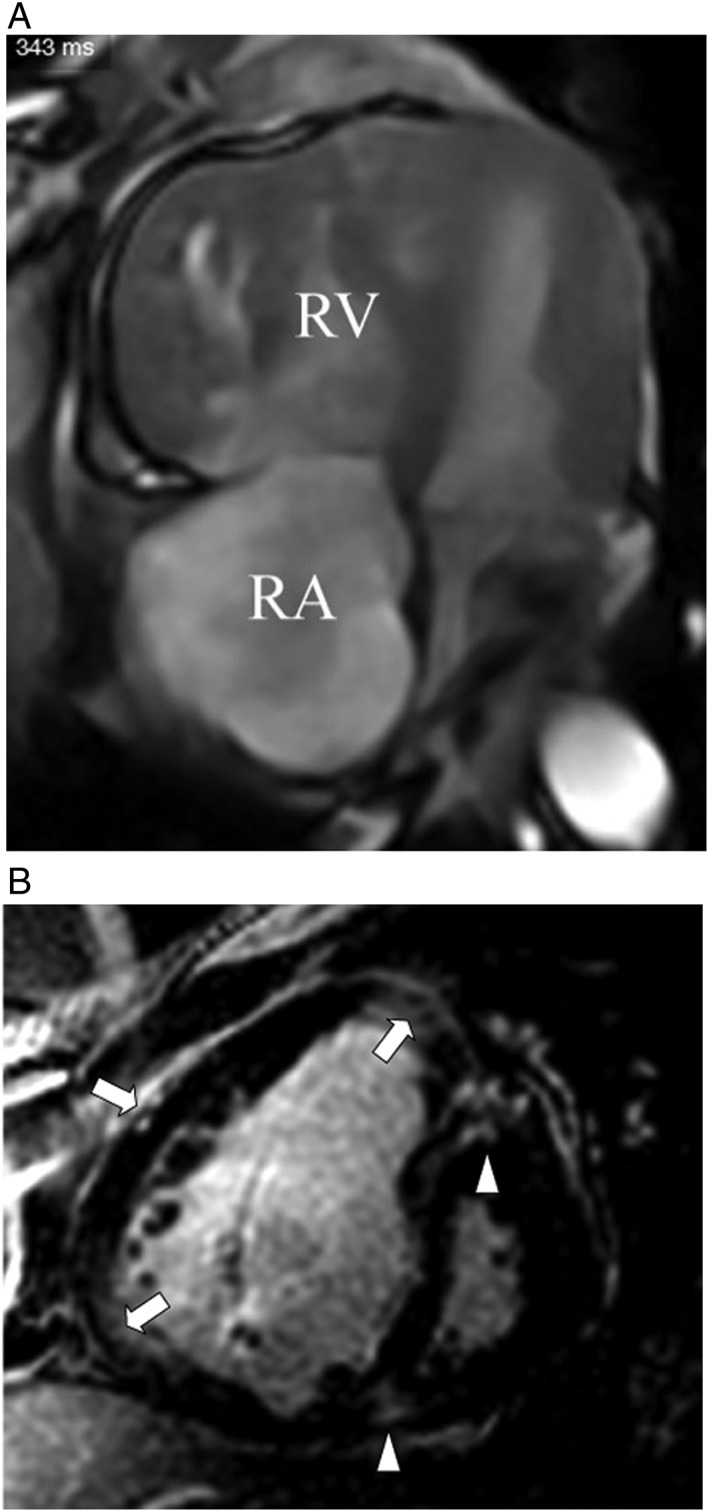
Figure 3.

(A) Magnetic resonance study (steady‐state‐free precession sequence, horizontal long axis view, and end‐diastolic frame) demonstrates dilation of the right ventricle (RV) and right atrium (RA), marked right ventricular hypertrophy, with displacement of the interventricular septum towards the left ventricle; both left ventricle and atrium are compressed. (B) Contrast‐enhanced magnetic resonance study (inversion recovery‐gradient echo sequence, short axis view, and end‐diastolic frame) of the identical patient with pulmonary vascular sarcoidosis and resulting severe pulmonary arterial hypertension demonstrates contrast‐enhancement of the right ventricular hinge points (triangles) and free wall (arrows).
