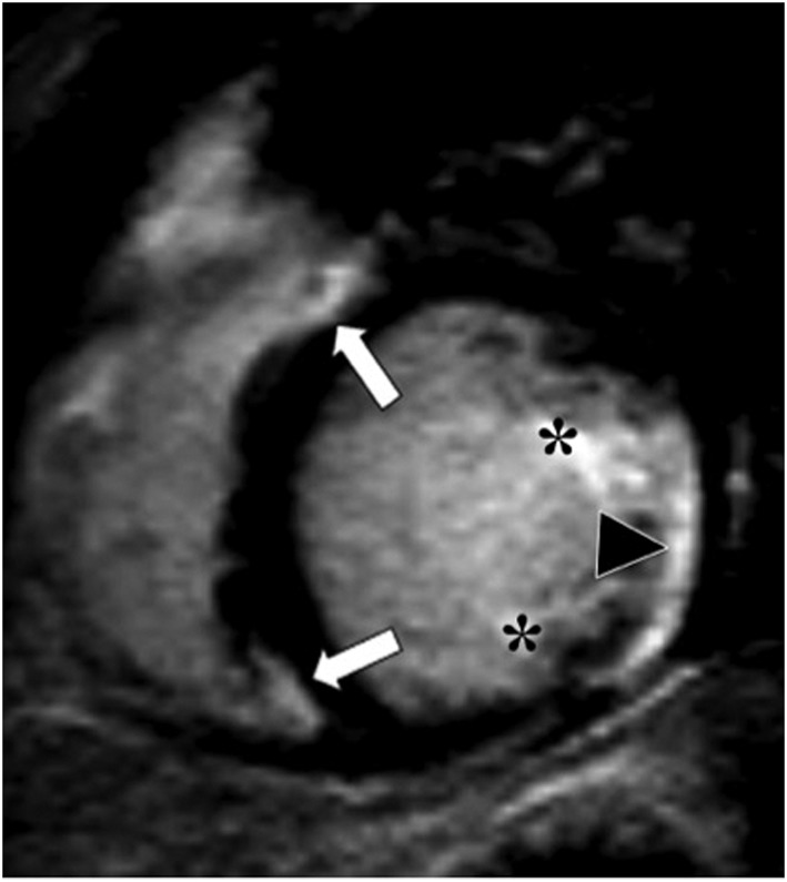
Figure 4.

Contrast‐enhanced magnetic resonance study (inversion‐recovery gradient echo sequence and short axis view) in a patient without pulmonary hypertension demonstrates enhancement of the ventricular insertion points (arrows), papillary muscles (asterisks), and postero‐lateral left ventricular segments (triangle).
