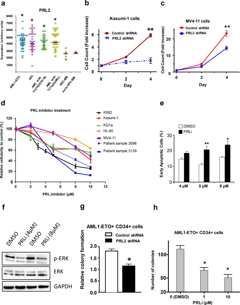
Figure 1.
PRL2 promotes the proliferation and survival of human AML cells. (a) PRL2 is highly expressed in some subtypes of human AML compared to normal human bone marrow HSPCs (*P<0.05). (b) Kasumi-1 cells were transduced with lentiviruses expressing control or PRL2 shRNA. The proliferation of transduced cells (GFP+) was measured over time (**p<0.01, n=3). (c) MV4-11 cells were transduced with lentiviruses expressing control or PRL2 shRNA. The proliferation of transduced cells (GFP+) was measured over time (**p<0.01, n=3). (d) Inhibiting of PRL2 activity with a small molecule PRL inhibitor (PRLi) decreases the viability of human AML cell lines and primary human AML cells in a dosage-dependent manner. Patient sample 3096 is from an AML patient positive for FLT3ITD and FLT3TKD mutations. Patient sample 3139 is from an AML patients negative for FLT3ITD and NPM mutations. (e) Human K562 cells were treated with PRL inhibitor (PRLi) for 24 hours and apoptosis was determined by Annexin V and DAPI staining (*p<0.05, **p<0.01, n = 3). (f) Immunoblot analysis of ERK phosphorylation in human K562 cells following DMSO or a small molecule inhibitor (PRLi) treatment. Representative Western blot analysis of indicated proteins is shown. (g) Human cord blood CD34+ cells expressing AML1-ETO were transduced with lentiviruses expressing control or PRL2 shRNA. Myeloid progenitors were quantified by using the methylcellulose culture (*p<0.05, n=3). (h) PRL inhibitor (PRLi) treatment decreases the colony formation of human cord blood CD34+ cells expressing AML1-ETO in a dosage dependent manner (*p<0.05, n = 3).

