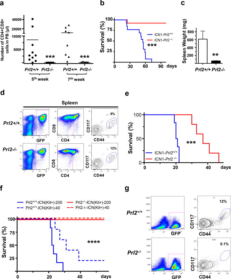
Figure 2.
PRL2 promotes oncogenic NOTCH1 induced T-ALL in vivo. (a) Lethally irradiated B6.SJL mice were transplanted with Prl2+/+ or Prl2−/− progenitor cells expressing NOTCH1-ICN1, and peripheral blood was collected and the frequency of donor-derived (GFP+) CD4+CD8+ cells was determined by FACS analysis (n=10, ***p<0.001). (b) Lin− bone marrow cells isolated from Prl2+/+ and Prl2−/− mice were transduced with retroviruses expressing NOTCH1-ICN1 and equivalent number of transduced cells (GFP+) were injected into lethally irradiated recipient mice. Kaplan-Meier curve shows the survival of mice transplanted with cells expressing NOTCH1-ICN1 for the period of observation (n=12, ***p<0.001). (c) Average spleen weight in recipient mice repopulated with Prl2+/+ or Prl2−/− cells expressing NOTCH1-ICN1 (n=10, **p<0.01). (d) Prl2+/+ and Prl2−/− fetal liver cells were transduced with retroviruses expressing NOTCH1-ICN1 and equivalent number of transduced cells (GFP+) were injected into lethally irradiated recipient mice. Representative flow cytometry blots showing the frequency of leukemia–initiating cells (Kit+CD44+CD25+) in the spleen of primary recipient mice 4 weeks following transplantation. (e) 20,000 GFP+ cells isolated from the spleens of primary recipient mice were injected into lethally irradiated recipient mice. Kaplan-Meier curve shows the survival of mice transplanted with cells expressing NOTCH1-ICN1 for the period of observation (***p<0.001, n=5). (f) 40 or 200 leukemia–initiating cells (Kit+CD44+CD25+) purified from the spleens of primary recipient mice were injected into lethally irradiated recipient mice. Kaplan-Meier curve shows the survival of mice transplanted with cells expressing NOTCH1-ICN1 for the period of observation (****p<0.0001, n=5). (g) Representative flow cytometry blots showing the frequency of leukemia–initiating cells (Kit+CD44+CD25+) in the spleens of secondary recipient mice repopulated with leukemia–initiating cells (Kit+CD44+CD25+) expressing NOTCH1-ICN1.

