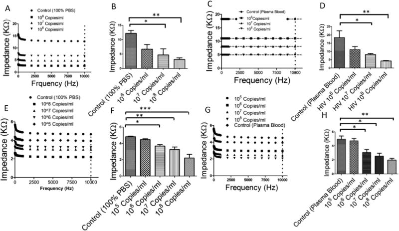
Fig. 4.
HIV-1 capture and detection using cellulose paper microchips with graphene-modified silver electrodes. (A–D) Virus capture and detection using on-chip physical antibody immobilization. (A) Impedance spectroscopy of viral lysate for frequencies between 1 Hz and 10 kHz. (B) Impedance magnitudes of viral lysate samples at 10 kHz. We were able to detect HIV-1 in spiked PBS samples with concentrations of 107 copies per ml and 108 copies per ml. (C) Impedance spectroscopy of viral lysate samples in HIV-spiked plasma samples. (D) Impedance magnitude of viral lysate in HIVspiked plasma samples at 10 kHz. (E–H) Virus capture and detection using the on-chip covalent immobilization of anti-gp120 antibody. (E) Impedance spectroscopy of viral lysate samples in HIV-spiked PBS. (F) Impedance magnitude of viral lysate samples in HIV-spiked PBS at 10 kHz. The impedance magnitude of viral lysate samples in HIV-spiked PBS with a viral load of 106 copies per ml was significantly different than virus-free control samples. (G) Impedance spectroscopy of viral lysate for HIV-spiked plasma. (H) Impedance magnitude of viral lysate for HIV-spiked plasma samples at 10 kHz. The impedance magnitude of the viral lysate of the HIV-spiked plasma with a viral load of 106 copies per ml was different than the virus-free control.

