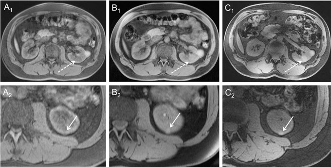
Fig 7. 3D VIBE.
3D VIBE imaging at 1.5 Tesla (A), 3 Tesla (B) and 7 Tesla (C) in the same subject. 7 Tesla 3D VIBE imaging demonstrated diagnostic potential by means of detection of pathologies, as it revealed a second hemorrhaged renal cyst (dashed arrow Figure C1), not being displayed at lower field strengths. In the second row, arrows show a further very small renal cyst in the same subject, which is also best visible at 7 Tesla (C2).

