
Figure 1.
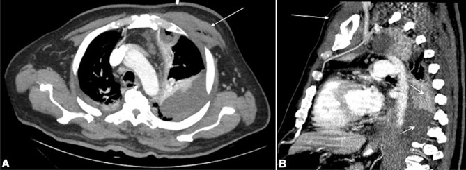
Thoracic CT scan (A) axial and (B) sagittal cuts showing pectoralis major pyomyositis (long arrows) and pleural empyema (short arrows) contacting the thoracic vertebrae.
Official websites use .gov
A
.gov website belongs to an official
government organization in the United States.
Secure .gov websites use HTTPS
A lock (
) or https:// means you've safely
connected to the .gov website. Share sensitive
information only on official, secure websites.
Thoracic CT scan (A) axial and (B) sagittal cuts showing pectoralis major pyomyositis (long arrows) and pleural empyema (short arrows) contacting the thoracic vertebrae.