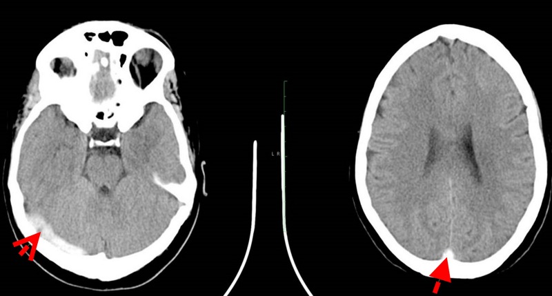
Figure 1.
Brain CT findings on admission. The first red arrow shows a left parasagittal parietal lobe high convexity gyral hypodensity (1.8×0.9 cm) region is seen. Appearances may be caused by a venous infarct. The second red arrow shows an abnormal superior sagittal and left sigmoid sinus hyperdensity suspicious for venous sinus thrombosis (empty delta sign). Relative hypodensity in the left internal capsule. No other areas of abnormal attenuation. Otherwise normal appearances of the brain parenchyma, ventricles, cisterns and nuclei. No extra-axial collections. No intraparenchymal haemorrhage detected. Impression: left parietal lobe venous infarct. Venous dural sinus thrombosis.

