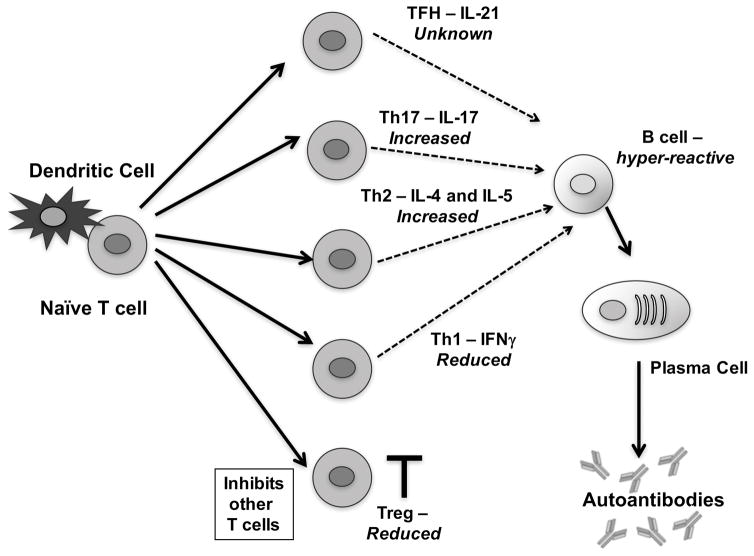
FIGURE 3. T cell aberrations in Ets1-deficient mice.
Schematic of T cell differentiation with major subsets that have been implicated in lupus shown. Alterations in T cell subsets in Ets1-deficient mice are indicated in italics. Naïve autoreactive T cells interact with antigen-presenting dendritic cells and subsequently differentiate into various T cell subsets, depending on the cytokine environment in which they become activated. T cells can differentiate into T follicular helper (TFH) cells, T helper 17 (Th17) cells, T helper 2 (Th2), T helper 1 (Th1) or regulatory T cells (Treg). Tregs function to inhibit the other T cell subsets and limit autoimmune disease. Tregs are reduced in Ets1-deficient mice. The other T cell subsets produce various cytokines (as shown in the figure) and can interact with B cells to promote proliferation, germinal center formation, isotype-switching, affinity maturation and plasma cell formation. Ets1-deficiency results in skewing towards Th17 and Th2 cytokines and away from Th1 cytokines, while the effect on TFH cells is unknown. Depending on the type of T cells that interact in vivo with B cells, differences in B cell outcomes are possible. B cells from Ets1 knockout mice are also hyper-responsive and have an intrinsic propensity to differentiate into plasma cells.

