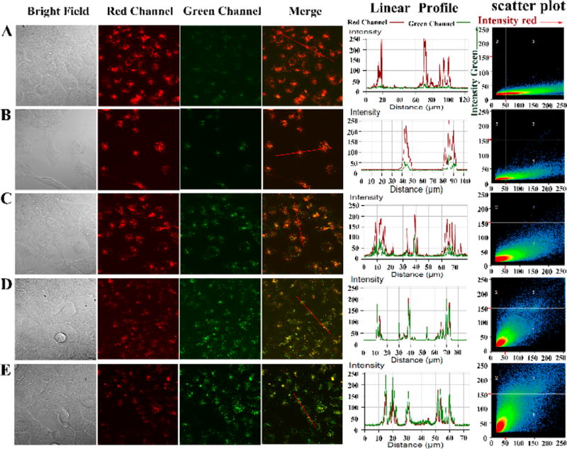
Figure 5.
Confocal fluorescence imaging of exogenous ClO− in MCF-7 cells. MCF-7 cells were first incubated with PFOBT36SeTBT5 Pdots (10 µg/mL) overnight at 37 °C and then were incubated with different solutions for 30 min: (A) PBS solution; (B) 10 µM NaClO; (C) 25 µM NaClO; (D) 50 µM NaClO; and (E) 80 µM NaClO. Fluorescence images were acquired using a confocal microscope with 405 nm excitation and fluorescence emission windows of green channel (530–600 nm) and red channel (>650 nm).

