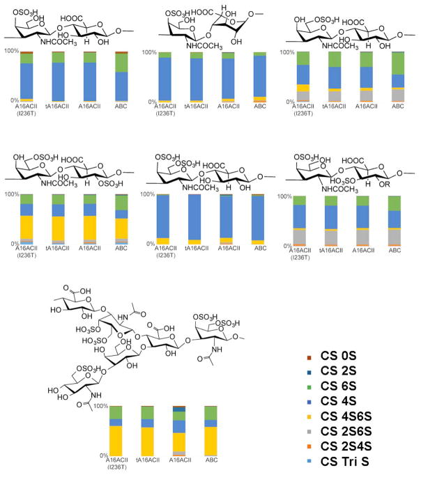
Figure 4.
The enzymes discussed in this study are 1: tA16ACII(I236T), 2: tA16ACII, 3: A16ACII, and 4: CS ABC) were tested against a panel of various CS substrates. Each bar chart shows the percent composition of disaccharide on the x-axis while the y-axis illustrates which enzyme is involved in the reaction. Each reaction consisted of two independent replicates that were pooled for LC-MS disaccharide analysis. Above each bar chart is the characteristic repeating disaccharide structure found in each substrate, comprising anywhere from 10% to 90% of each structure. The structure of CS-E from M. chinensis is based on the work of Higashi et al. [11]. The structure of octopus CS-K is currently unavailable.

