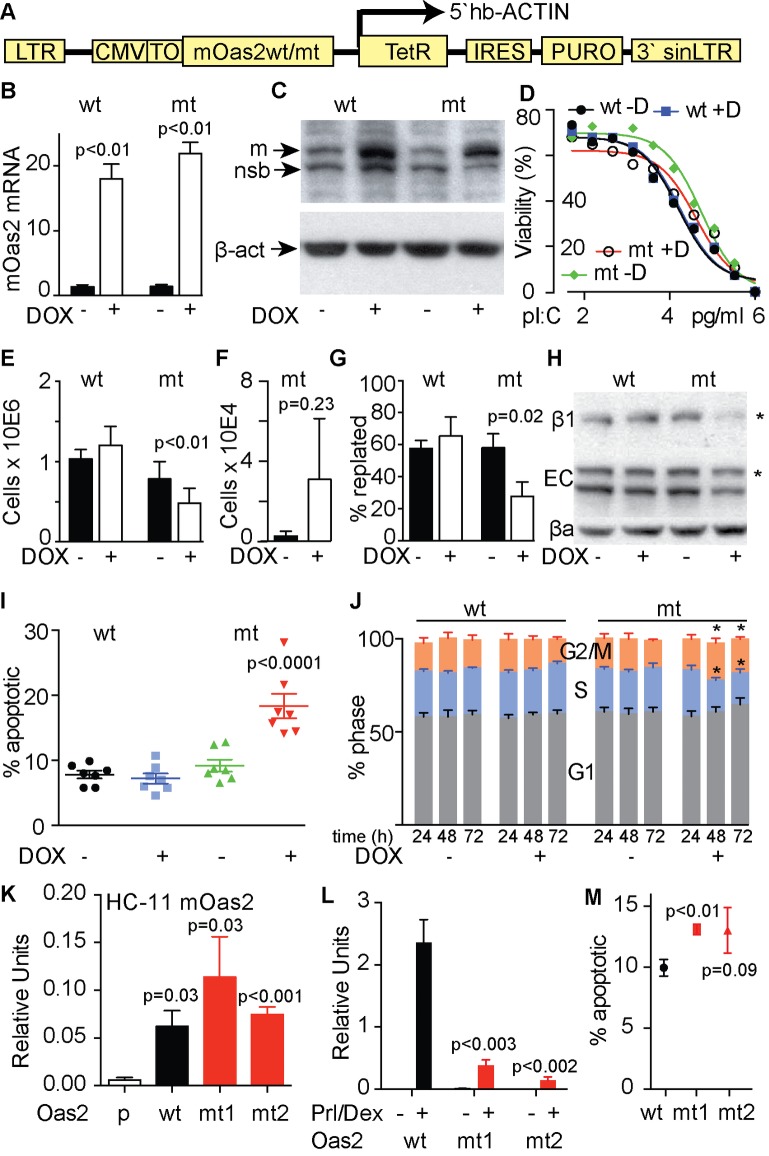
Fig 4. The effects of inducible expression of mutant and wild type Oas2 in T47D cells.
(A) pHUSH ProEx expression vector used to express either mutant (mt) or wild type (wt) mouse Oas2 in T47D cells in response to doxycycline (DOX). (B) relative expression of mt and wt Oas2. (C) Western blot showing induction of mouse OAS2 (m) running just below endogenous human OAS2 protein, with both bands above a non-specific band (nsb). (D) Sensitivity of the cells lines to poly I:C (pl:C) with and without DOX induction of mt and wt Oas2. (E) Effect of mt and wt Oas2 on adherent cell number after 72h. (F) Cell detachment (numbers of live cells in supernatant fraction) caused by mt Oas2. (G) Effects of mt or wt Oas2 on replating of T47D cells in a 4 hour trypsin only replating assay after 48h of DOX. (H) Expression of β1 integrin (β1), E-cadherin (EC) and β-actin (βa) in response to induction of mt or wt Oas2. (I) apoptotic response to induction of mt or wt Oas2. Data represents the average of 7 independent experiments. (J) cell-cycle-phase distribution at the indicated times following induction of mt or wt Oas2. Data represents the average of 5 independent experiments. *p<0.01. ANOVA 4I and J. (K) Oas2 expression in parental (p) normal mouse mammary HC11 cells or in cells constitutively expressing mt or wt Oas2. (L) Effect of wt or mt Oas2 on beta Casein in HC11s after 72 hours of prolactin (Prl) and Dexamethasone (Dex) stimulation. (M) Effect of mt or wt Oas2 expression on cell death at 96 hours in HC11 cells after transient transfection. All data are representative of 3 independent experiments in response to 72h of DOX except otherwise specified. Paired t-tests 4B,E,F, G, L and M.

