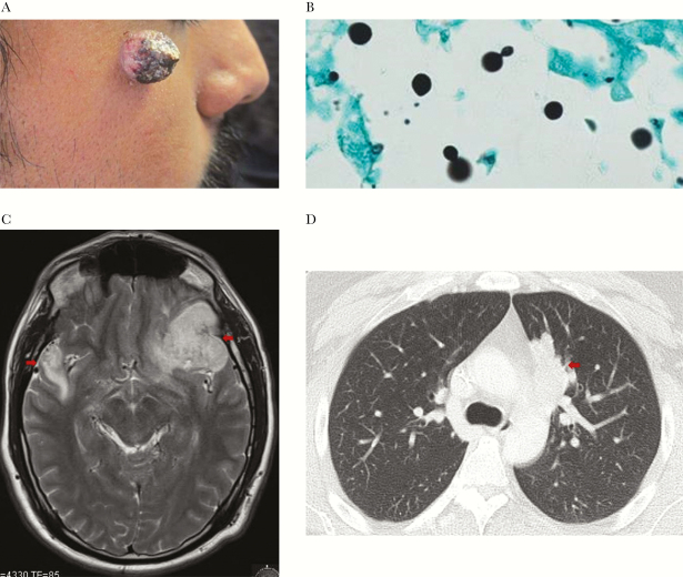
Figure 3.
(A) Right face skin lesion due to cutaneous Cryptococcus gattii.(B) Skin biopsy, Gomori methenamine silver stain with 100× magnification with narrow based budding yeast consistent with Cryptococcus. (C) Magnetic resonance image of the brain showing irregular rim-enhancing intracranial masses involving the left frontal lobe and right temporal lobe (arrows). (D) Computed tomography scan of the chest with a 4.7-cm mass (arrow) in medial aspect of the left upper lobe.

