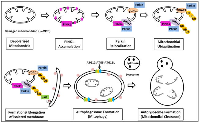
Figure 3.
Mitophagy: Summary of steps involved in mitophagy. The depolarized mitochondrion leads to accumulation of PINK1, which phosphorylates Mfn2, which acts as a lure for Parkin. Parkin binding Mfn2 triggers mitophagy. Outer membrane proteins, including both Mfn’s and VDAC, are ubiquitinated. P62 is recruited, binding the ubiquitinated proteins, linking them to LC3. The isolation membrane elongtes and eventually engulfs the mitochondrial pieces destined for autophagy, forming the autophagosome, which eventually fuses with the lysosome, leading to degradation of the enclosed mitochondria.

