Figure 3.
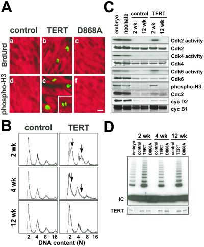
Cardiac myocyte DNA synthesis and mitoses in αMHC-TERT transgenic mice. (A) Immunofluorescence detection of BrdUrd incorporation (green; a–c) and mitotic phosphorylation of histone H3 (green; d–f) in cardiac myocytes at 2 wk of age, identified with MF20 Ab to sarcomeric MHC (red). Neither BrdUrd incorporation nor histone H3 phosphorylation was detected in nontransgenic controls or D868A mice. A cardiac myocyte in late telophase is seen in e (Inset). [Bar =10 μm.] (B) Dissociated cardiac myocytes were analyzed by flow cytometry. Arrows highlight the increase in DNA content by TERT. (C) TERT prolongs the activity of endogenous Cdk6, Cdk2, and Cdc2 in myocardium. Cdk2/4/6 activities were monitored by immune complex kinase assays, and Cdc2 activity was monitored by Ser-10 phosphorylation of histone H3. Western blots indicate an increase in cyc D2, cyc B1, and Cdc2 but not other Cdks. (D) The shift from hyperplasia to hypertrophy does not entail down-regulation of TERT expression (Western blot Bottom) or activity (TRAP assay Top).

