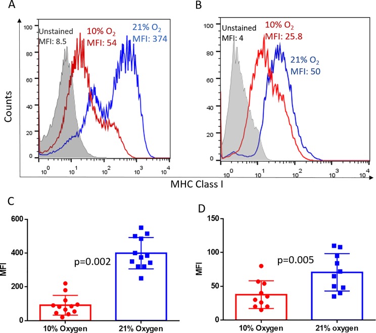
Fig 1. Hypoxia downregulates MHC class I expression on tumor cells in vivo.
Whole body exposure of mice with 11 day established MCA205 pulmonary tumors (A, C; n = 6 per group per experiment) or 15 day established subcutaneous tumors (B, D; n = 5 per group per experiment) to 10% oxygen for 48h significantly downregulated MHC class I expression on tumor cells as compared with mice breathing 21% oxygen. MHC class I levels were determined by flow cytometry. Representative histograms (A and B) and associated quantification and statistics (C and D) of 2 independent experiments are shown. The significance of differences was analyzed by the Student’s t-test (two-sided); p = 0.002 (C), p = 0.005 (D). Grey filled: Unstained control; Red: Hypoxia; Blue: Normoxia. MFI: mean fluorescence Intensity. Error bars indicate SD.

