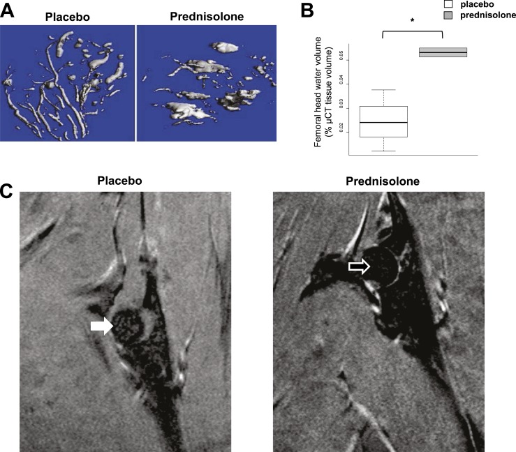
Figure 9.
Femoral head vascularity after implantation of placebo or 14 days of prednisolone administration. (A) µCT after lead chromate infusion in the femoral head shows that prednisolone transforms the normal dendritic vascularity of the femoral head to large pools of edema. (B) Femoral head water volume. *P < 0.05 (n = 3). Water restrained or bound by vessels changed to more mobile free edema. (C) MRI obtained with T1 weighting reveals a higher image intensity ratio of the femoral head in the animal receiving prednisolone (open arrow, 0.334) than in the animal receiving placebo (closed arrow, 0.313), indicative of more free water or edema.

