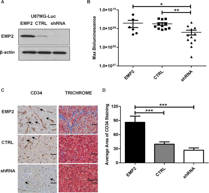
FIG 1. EMP2 Promotes Angiogenesis in U87MG Intracranial Tumors.
(A) EMP2 levels were validated among the U87MG/Luc panel by Western Blots prior to stereotactic implantation. (B) Intracranial tumor growth was monitored by bioluminescence imaging on day 18 post tumor implantations in mice bearing U87MG/EMP2 (n=6), U87MG/CTRL (n=11) or U87MG/shRNA (n=12). The numbers of animals per group was generated by pooling two independent trials. (C) Intracranial tumors were fixed and stained with CD34 (left) and trichrome (right) and representative images were taken with a bright field microscope under 400× magnification. Arrowheads indicate representative staining of tumor associated vasculature. (D) Automated quantification of CD34 staining among groups was determined by NIH Image J software with a custom macro script as detailed in methods. One-way analysis of variance with a Bonferroni post-test was calculated to determine the difference among three groups. Significance was defined as *p<0.05, ** p<0.001, *** p<0.0001.

