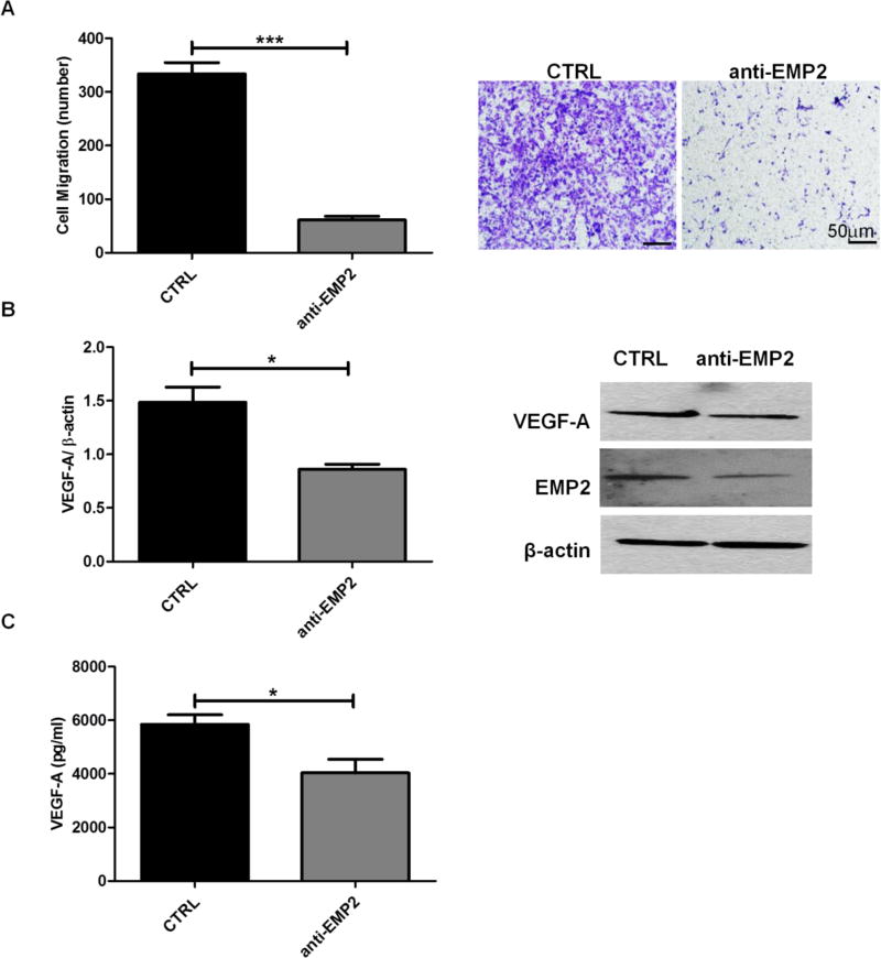
FIG 5. Anti-EMP2 Decreases HUVEC Migration and VEGF-A Expression and Secretion.
U87MG wild type cells were cultured in the presence of anti-EMP2 antibody (n=4) or a vehicle (saline) control (n=3) for 48 hours. (A) Conditioned media were collected at the end of incubation for HUVEC migration assay. Migratory cell numbers were averaged by counting four random fields per transwell (left) and representative images of migratory cells were shown under 400× magnification (right). (B) Quantification of VEGF-A expression normalized by β-actin were determined by NIH Image J software (left) and representative images of Western Blots show the expression of VEGF-A, EMP2, or β-actin (right). (C) Quantification of cell-secreted VEGF-A levels were determined by ELISA. The experiments were repeated at least three times. Student’s t test was used to determine the difference between the two groups. Significance was defined as *p<0.05, *** p<0.0001.

