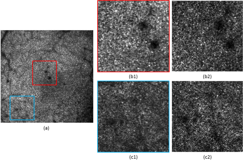
Fig. 7.
Comparison of the AO-SLO image quality between a large FoV and small FoV configuration, for healthy volunteer V2. (a) Image obtained on a 4°×4° FoV at the fovea. (b1) 1°×1° zoom from (a), at the fovea. (b2) Same area recorded with a 1°×1° FoV. (c1) 1°×1° zoom from (a), at 2.1° eccentricity from the fovea. (c2) Same area recorded with a 1°×1° FoV. The same grey scale is used for each image.

