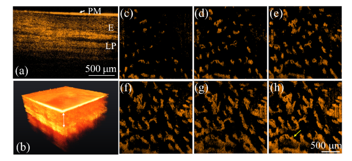
Fig. 7.
In vivo OCT images of gingival mucosa obtained from a 24-year-old female including a (a) 2D image, (b) 3D image, and (c)–(h) projection-view angiographies obtained at the various depths of 250–260, 300–310, 350–360, 400–410, 450–460, and 250–500 μm beneath the tissue surface. E: epidermis, LP: lamina propria, and PM: plastic membrane. The scale bar in (a) represents 500 μm.

