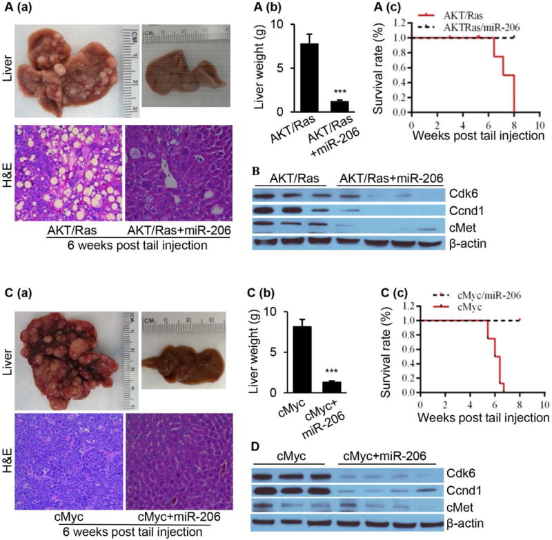
Fig. 5.

HCC was undetectable in the livers of both AKT/Ras and cMyc HCC mice after miR-206 overexpression. (A) (a) Macroscopic (upper panel) and microscopic (lower panel) appearance of livers from AKT/Ras/pT3 mice (control, n=10) and AKT/Ras/miR-206 mice (n=10) stained with H&E (100×); (b) Average liver weight of AKT/Ras/pT3 mice and AKT/Ras/miR-206 mice (n=10); and (c) Kaplan Meier survival curves of AKT/Ras/pT3 and AKT/Ras/miR-206 mouse cohort. (B) Western blot analysis of Cdk6, Ccnd1 and cMet in the livers of AKT/Ras and AKT/Ras/miR-206 mice. (C) (a) Macroscopic (upper panel) and microscopic (lower panel) appearance of livers from cMyc/pT3 mice (n=10) and cMyc/miR-206 mice (n=10) stained with H&E (100×); (b) Average liver weight of cMy/pT3 mice and cMyc/miR-206 mice; and (c) Kaplan Meier survival curve of cMyc/pT3 and cMyc/miR-206 mouse cohort. (D) Western blot analysis of Cdk6, Ccnd1 and cMet in the livers of cMyc and cMyc/miR-206 mice. Data represent mean ± SEM. ***p < 0.001.
