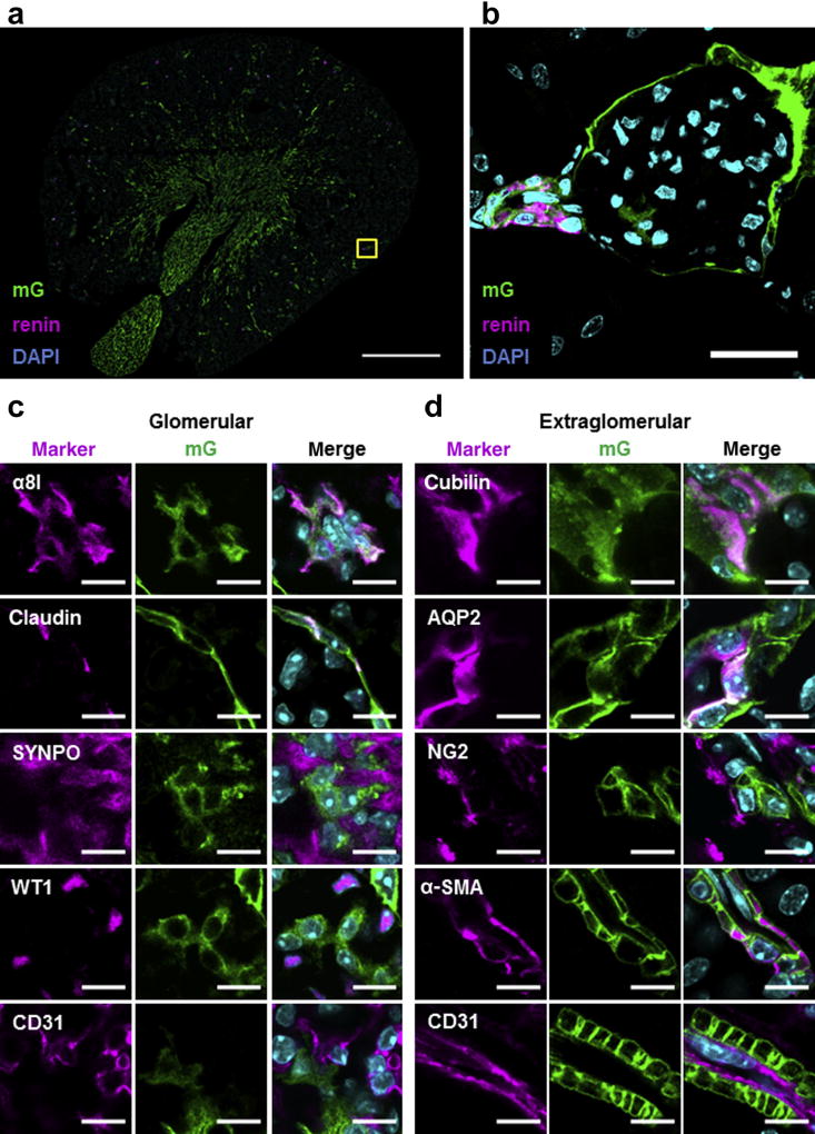
Figure 1. Renin lineage cells (RLCs) give rise to many different cell types in adult mouse kidneys.
(a) Representative overview image of RLCs (mG+) and their renin-positive subpopulation in the kidneys of adult mRenCre-mT/mG mice. Bar = 1 mm. (b) Renin-producing cells at their classical juxtaglomerular position (high-magnification confocal image of the yellow frame area indicated in [a]). The 4′,6-diamidin-2-phenylindol (DAPI); nuclear marker. Bar = 25 µm. (c) Representative confocal images of glomerular cell marker expression in RLCs; α8 integrin (α8I)-mesangial cell marker, claudin-parietal epithelial cell (PEC) marker, synaptopodin (SYNPO), Wilm’s tumor protein 1 (WT1)-podocyte cell markers, and cluster of differentiation (CD)31-endothelial cell marker. Merged images contain 3 fluorescent channels (for the corresponding cell marker, mG, and DAPI). Bar = 10 µm. (d) Representative confocal images of extraglomerular cell marker expression in RLCs; cubilin and aquaporin 2 (AQP2)-tubular cell markers (for the proximal tubule and collecting duct, respectively), NG2-interstitial cell marker, α-smooth muscle actin (α-SMA), and CD31-vascular cell markers (for vascular smooth muscle cells and endothelial cells, respectively). Merged images contain 3 fluorescent channels (for the corresponding cell marker, mG, and DAPI). Bar = 10 µm. To optimize viewing of this image, please see the online version of this article at www.kidney-international.org.

