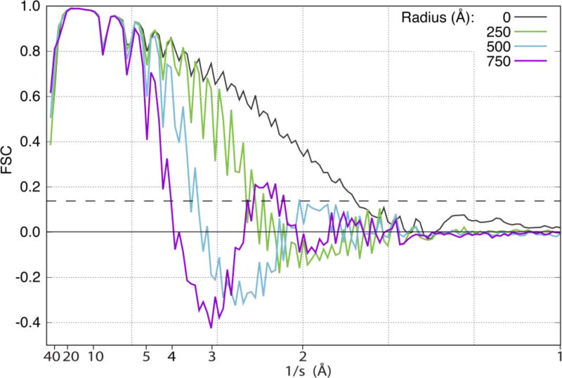
Fig. 6.
FSC curves for simulations that compare the maps for (1) a larger portion of the structure of tubulin located at different values of the radius, R, with (2) the density calculated directly from the coordinates in PDB ID: 1JFF. Results shown here are for maps located at radii of 0, 250, 500, and 750 Å. The dashed horizontal line is drawn at FSC = 0.143.

