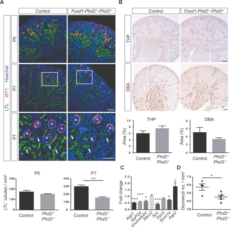
Figure 3. Combined inactivation of Phd2 and Phd3 in FOXD1 stroma reduces nephron formation.

(A) Shown are representative images of Lotus tetragonolobus lectin (LTL) staining (green) and IHC staining for Wilms tumor 1 (WT1) (pink); kidney sections were obtained from Cre− littermate control or Foxd1-Phd2−/−-Phd3−/− mutants at P0 or P7. Nuclei were stained with Hoechst dye. Arrows indicate proximal tubules and asterisks depict glomeruli. Scale bars: 100 μm (top and middle panels) and 50 μm (bottom panel). LTL+ tubules were quantified (n=3 each) for P0 and P7. (B) Shown are representative images of IHC staining for Tamm-Horsfall protein (THP) and Dolichos biflorus agglutinin (DBA) lectin staining of kidney sections from Cre− littermate controls and Foxd1-Phd2−/−-Phd3−/− mice at P7. THP+ and DBA+ tubules were quantified (n=3–5). Scale bar: 200 μm. (C) Fold changes in mRNA expression levels of nephron segment-specific genes in total kidney homogenates fromFoxd1-Phd2−/−-Phd3−/− mutants compared to Cre− littermate controls (n=8 each). (D) Quantification of glomerular numbers. Shown are number of glomeruli per mm2 (n=4–5). Data are represented as mean ± SEM; 2-tailed Student’s t-test, *p<0.05, **p<0.01 and ***p<0.001. Acta 2, α-smooth muscle actin; Aqp1, aquaporin 1; Aqp2, aquaporin 2; NaPi2a, sodium-phosphate co-transporter-2a; Ncc NaCl co-transporter; Nkcc2, Na-K-2Cl co-transporter, Scnn1a, sodium channel epithelial 1 alpha subunit; Trpv5, transient receptor potential cation channel subfamily V member 5.
