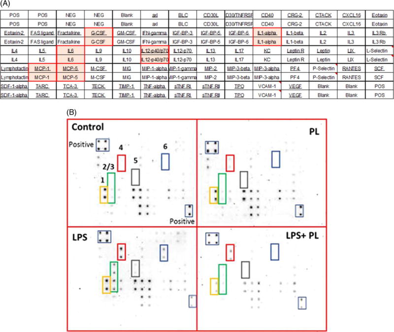
Fig. 4.

Plumbagin effect on different cytokine expression in LPS-activated BV-2 microglial cells. (A). Microarray layout AAM-CYT-3 was used to assess chemokines/cytokines expression in cell-free supernatant. (B). Microarray chemiluminescence detection. Four blots represented the supernatants of the following: control resting cells treated with DMSO, cells treated with 2 μM of PL, cell treated with LPS (1 μg/mL) and finally supernatant of LPS-activated cells after exposure to PL. The most affected cytokines are designated as follows: 1, MCP-1; 2, IL-6; 3, MCP-5; 4, G-CSF; 5, IL-12 p40/p70; 6, IL-1α.
