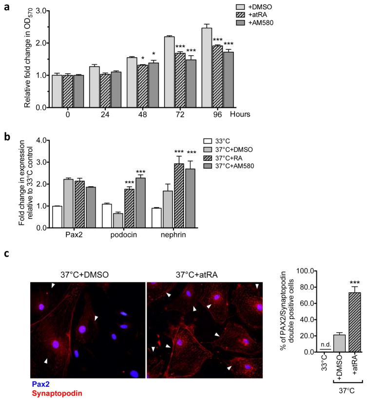
Figure 5. RA inhibits proliferation of PECs and enhances expression of podocyte marker synaptopodin in vitro.
(a) Immortalized mouse PECs were cultured in 37°C without IFN-γ for 14 days to induce their differentiation and were incubated with either vehicle (DMSO), atRA (5μM), or AM580 (200nM) for 96 hours. Crystal violet cell proliferation assay was performed to determine the effect of RA on PECs. Growth curve of differentiated PECs are shown as relative fold change in crystal violet stain (OD570nm) compared to 0h (no factors added) (*P<0.05 and ***P<0.001 compared to DMSO control; n=6). (b) Immortalized mouse PECs were cultured either at 33°C with IFN- γ or 37°C without IFN- γ to induce differentiation for 14 days. Following 14 days, cells were additionally treated with either vehicle (DMSO) atRA (5μM), or AM580 (200nM) for 72 hours. Real-time PCR analysis for PEC marker, Pax2, and podocyte markers, podocin, nephrin and synaptodopodin, are shown (*P<0.05, **P<0.01 and ***P<0.001 compared to 33°C-grown undifferentiated controls; ##P<0.01 and ###P<0.001 compared to DMSO-treated differentiated PECs. n=6). (c) Representative image of Pax2/synaptopodin immunofluorescence in differentiated PECs treated with vehicle or atRA (5μM) for 72 hours. Quantification of Pax2/synaptopodin co-expressing PECs/field is shown on the right (***P<0.001 compared to DMSO control).

