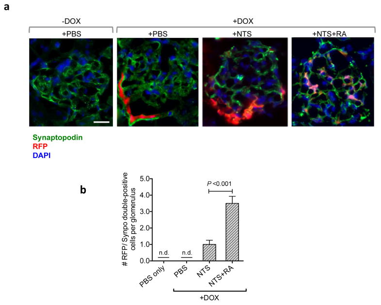
Figure 6. Lineage tracing of PECs shows increased PECs expressing podocyte marker synaptodopodin in vivo following RA admnistration in NTS-GN mice.
PEC-rtTA; TetO-Cre; CAGs-tdTomato transgenic mice were fed with DOX for 4 weeks before injection of NTS to label PECs with tdTomato RFP. (a) Representative images of glomeruli in mice with or without DOX induction show RFP-labeled PECs only with the DOX induction (magnification: x400, scale bar 20μM). RA treatment induced migration of PECs into glomerular tufts in NTS-treated mice. Co-localization of RFP with synaptopodin (in green) confirms the transdifferentiation of PECs into podocytes in NTS+RA glomeruli. (b) Quantification of RFP/synaptopodin (synpo)-double positive cells/glomerular cross section (n.d., not detected, n=6 per group).

