
Figure 3.
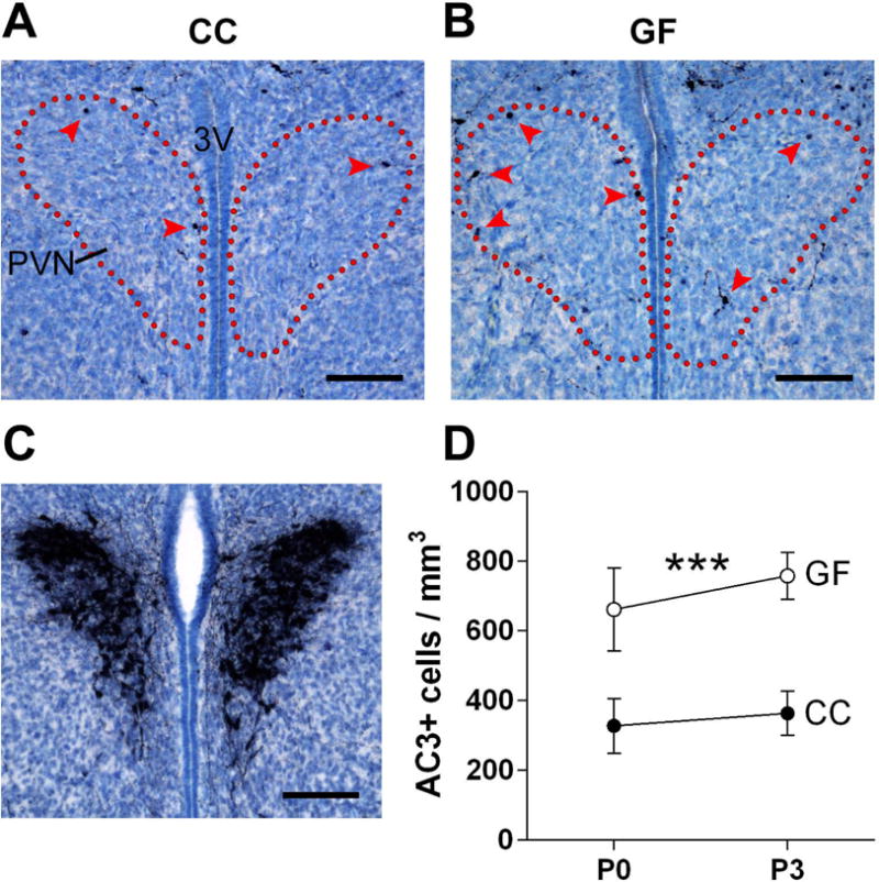
The absence of a microbiota increases cell death in the neonatal mouse paraventricular nucleus of the hypothalamus (PVN). (A, B) Photomicrographs of AC3-labeled tissue (counterstained with thionin) in representative conventionally colonized (CC) and germ-free (GF) mice on the day of birth. Dotted lines indicate the outlines of the PVN and arrowheads indicate AC3 labeled cells. 3V, third ventricle. Scale bars = 100 μm. (C) Vasopressin immunoreactivity was used to confirm the location of the PVN in the neonatal mouse brain. Scale bar = 100 μm. (D) Cell death density in CC (filled circles) and GF (open circles) mice at postnatal day (P) 0 and P3. Asterisks represent main effect of microbiota status. *** p < 0.001. N = 10 – 12 mice per group.