
Figure 4.
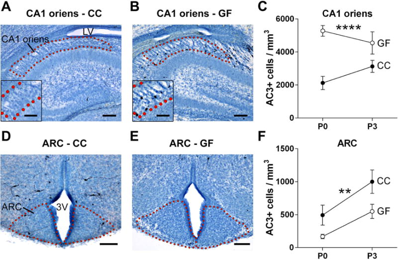
The absence of a microbiota alters developmental cell death in subregions of the hippocampus and hypothalamus. (A, B, D, and E) Photomicrographs of AC3-labeled tissue (counterstained with thionin) in sections containing the CA1 oriens layer of the hippocampus and arcuate nucleus of the hypothalamus (ARC) of conventionally colonized (CC) and germ-free (GF) mice on postnatal day (P) 0. Dotted lines outline the regions of interest; insets are a higher magnification view of the region shown above. 3V, third ventricle; LV, lateral ventricle. Scale bars = 100 μm (A,B,D,E) and 50 μm (insets A, B). (C, F) Cell death density in the CA1 oriens and ARC in CC (filled circles) and GF (open circles) mice at P0 and P3. Asterisks represent main effect of microbiota status. ** p < 0.01; **** p < 0.0001. N = 10 – 12 mice per group.