
Figure 6.
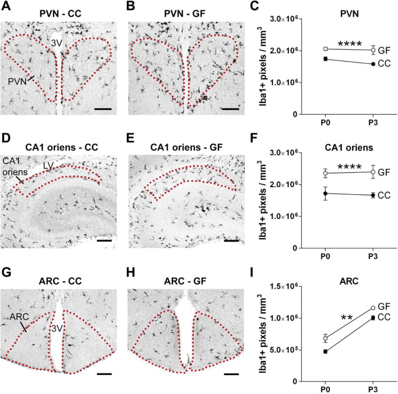
The absence of a microbiota causes increased microglial labeling in subregions of the hypothalamus and hippocampus in the neonatal mouse brain. (A, B, D, E, G, H) Photomicrographs of Iba1 labeled sections containing the PVN (A, B), CA1 oriens (D, E) and ARC (G, H) in conventionally colonized (CC) and germ-free (GF) mice at postnatal day (P) 0. Dotted lines outline the regions of interest. 3V, third ventricle; LV, lateral ventricle. Scale bars = 100 μm. (C, F, I) Microglial labeling density in the PVN, CA1 oriens, and ARC in CC (filled circles) and GF (open circles) mice at P0 and P3. Error bars are smaller than symbols for the GF group in C and I. Asterisks represent main effect of microbiota status. ** p < 0.01; **** p < 0.0001. N = 913 mice per group, except N = 5 mice for GF-P3.