
Figure 7.
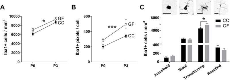
The absence of a microbiota alters the number, size and morphology of microglia in the neonatal mouse paraventricular nucleus of the hypothalamus (PVN). (A) Total microglia density in conventionally colonized (CC; filled circles) and germ-free (GF; open circles) at postnatal day (P) 0 and P3. (B) Average size of microglia in the PVN (in pixels). Asterisks represent main effect of microbiota status (A,B). (C) Top: Photomicrographs of amoeboid, stout, transitioning, and ramified microglia. Scale bar = 20 μm. Bottom: Density of microglial phenotypes in CC (black bars) and GF (grey bars) at P0 and P3 (ages combined for this analysis). Asterisk represent effect of microbiota status on transitioning microglia. * p < 0.05 and *** p = 0.0004. N = 11 – 13 mice per group except N = 6 mice GF-P3 (A,B); N= 18 – 24 mice per group (C).