
Figure 8.
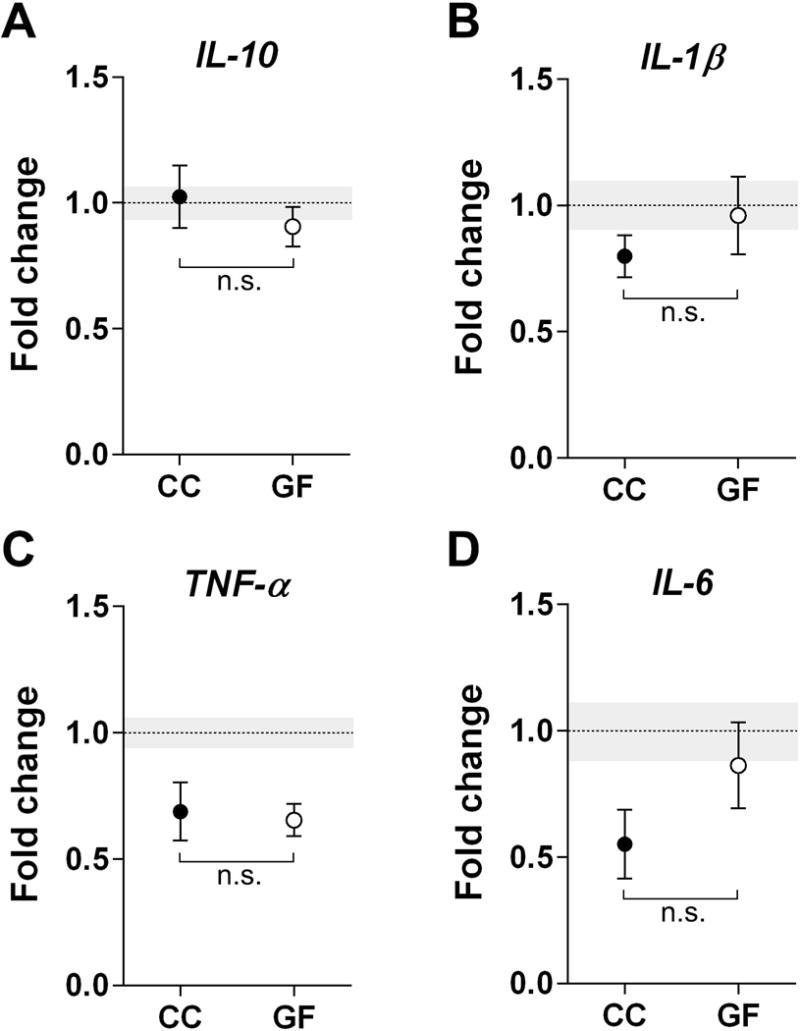
The absence of a microbiota does not affect cytokine expression in the prenatal mouse brain. Expression levels of anti-inflammatory IL-10 (A) and pro-inflammatory cytokines, IL-1β, TNF-α, and IL-6 (B-D), did not differ between conventionally colonized (CC; filled circles) and germ-free (GF; open circles) embryos. Data are expressed relative to levels of CC mice on P0. The mean of the control group is represented by a dotted line and its SEM by grey shading. n.s., non-significant. N = 12 –21 mice per group for (A); N = 12 – 22 mice per group for (B); N = 820 mice per group for (C); N = 7 – 14 mice per group for (D).