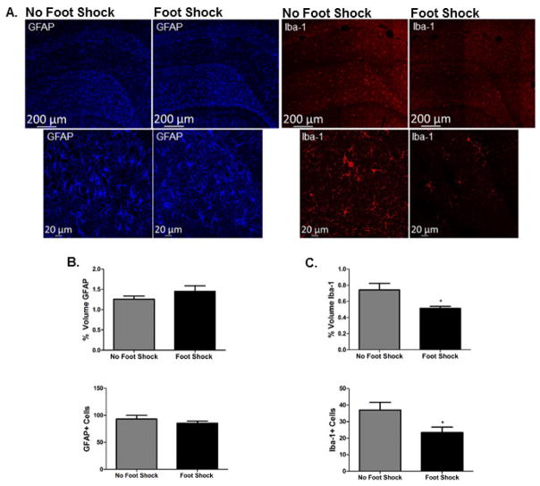
Figure 3. Dorsal hippocampal Iba-1 immunoreactivity, but not GFAP immunoreactivity, is attenuated 48 hours after severe stress.
A. Representative images of GFAP and Iba-1 immunoreactivity acquired at 10X (tiled image presented) and 20X are shown from stressed (Foot Shock in context A) and non-stressed (No Foot Shock in Context A) rats. Images were acquired in the DH, AP −3.36 from bregma. B. Both Imaris quantification and individual GFAP-positive cell counts indicated there was no effect of foot shock on GFAP immunoreactivity. C. In contrast, Imaris quantification and individual Iba-1 positive cell counts revealed that stress exposure significantly attenuated Iba-1 immunoreactivity 48 hours post-stress. * p < 0.05.

