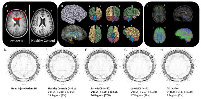
Figure 1.
A) MRI of Patient IH relative to an age-matched healthy control subject; B) cortical parcellation and volumetric modeling; C) diffusion imaging tractography; D–H) group-specific connectograms for Patient IH, Healthy Older Adults, Early MCI, Late MCI, and AD, respectively, with accompanying “Goodness-of-Fit” assessment of Patient IH’s group membership.

