Figure 2.

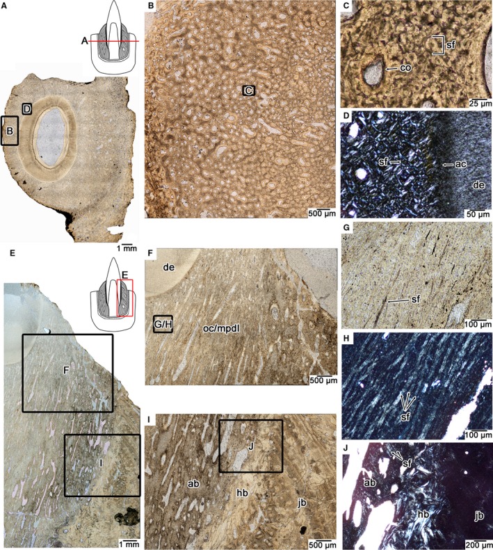
Tooth attachment tissue histology in the mosasaurid Platecarpus. (A) Wholeview of a transverse section through a single tooth root (UALVP 57045). (B) Closeup image of the tissue previously referred to as ‘bone of attachment’ (Rieppel & Kearney, 2005) or osteocementum (Caldwell et al. 2003) that fuses the tooth to the surrounding jawbone. (C) Closeup of (B) showing the fibrous microtexture of the attachment tissue. Most of the matrix is composed of Sharpey's fibers that extend parallel to the long axis of the tooth and are therefore circular in cross‐section. (D) Closeup of the attachment tissue surrounding the tooth root under cross‐polarized light, highlighting its fibrous microtexture. (E) Wholeview image of a Platecarpus dentary tooth in coronal section (UALVP 57044). (F) Closeup of (E) tooth attachment tissues in coronal view. (G) closeup of attachment tissue in coronal section under plain and cross‐polarized light (H) showing the network of Sharpey's fibers constituting the attachment tissue. (G and H) are taken from the same location. (I) Interface between the mineralized periodontal ligament, alveolar bone, remodelled attachment tissues, and jawbone. (J) Closeup image of alveolar bone and underlying remodelled attachment tissues. Note the presence of Sharpey's fibers, which mark the anchoring points for the periodontal ligament into the alveolar bone. ab, alveolar bone; ac, acellular cementum; co, cementeon; de, dentine; hb, Haversian bone; jb, bone of the jaw; mpdl, mineralized periodontal ligament; oc, osteocementum; sf, Sharpey's fibers.
