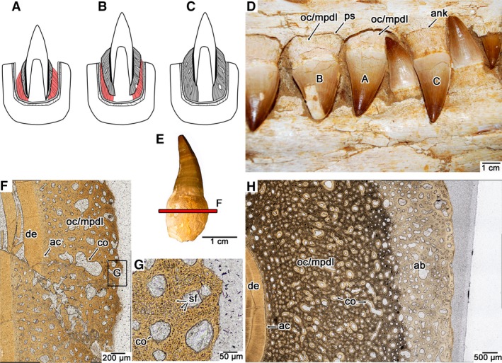
Figure 3.

Ontogeny of tooth attachment in mosasaurids. (A) Early stage gomphosis in an erupting tooth, where cementum (darker grey) and alveolar bone (lighter grey) are incipiently developed, but must be attached by PDL (red). (B) Later stage gomphosis in an erupted tooth, where cementum (darker grey) is continuing to mineralize centrifugally, calcifying the PDL (red). (C) Ankylosis in an erupted tooth, where cementum (darker grey) meets the alveolar bone (lighter grey), resulting in a completely calcified PDL. (D) Image of a set of maxillary teeth from the holotype of the mosasaurid Eremiasaurus (UALVP 51744) at the three stages of dental ontogeny (specimen was not used for thin sectioning). (E) Isolated tooth of Platecarpus with an intact root (UALVP 53595). These types of teeth were not shed and must have fallen out of the jaws postmortem, due to decay of the PDL. (F) Closeup image of a transverse section through an isolated tooth root of Platecarpus (UALVP 57046) showing the early development of the cementum (which is mainly composed of Sharpey's fibers of the PDL, and thus is also termed the mineralized PDL here). (G) Closeup image of the external root surface in the cross‐section figured in (F), showing Sharpey's fibers entombed in the osteocementum. (H) Closeup image of an ankylosed tooth of Platecarpus (UALVP 57045) showing the completed centrifugal calcification of the PDL. ab, alveolar bone; ac, acellular cementum; ank, ankylosis; co, cementeon; de, dentine; mpdl, mineralized periodontal ligament; oc, osteocementum; ps, space for the periodontal ligament; sf, Sharpey's fibers.
